Différences entre les versions de « Human model »
(Page créée avec « == Head == THE HEAD and more specifically the brain is among the most vital organs of the human body. However, these are not adapted to the dynamical loading conditions i... ») |
(→Head) |
||
| Ligne 6 : | Ligne 6 : | ||
From a mechanical point of view, the biological evolution of the head has lead to a number of integrated protection devices. The scalp and the skull but also to a certain extent the pressurized sub arachnoidal space and the dura matter are natural protections for the brain. | From a mechanical point of view, the biological evolution of the head has lead to a number of integrated protection devices. The scalp and the skull but also to a certain extent the pressurized sub arachnoidal space and the dura matter are natural protections for the brain. | ||
| + | |||
| + | === Adult FEM === | ||
| + | ==== Presentation ==== | ||
| + | The Strasbourg University Finite Element Head Model (SUFEHM) model was developed by Strasbourg University in 1997. | ||
| + | |||
| + | Figure shows a cross section of the model and illustrates the main anatomical features that are represented in the model which include the skull, falx, tentorium, subarachnoid space, scalp, cerebrum, cerebellum, and the brainstem. | ||
| + | |||
| + | The geometry of the inner and outer surfaces of the skull were digitized from a human adult male skull. | ||
| + | Data given in an anatomical atlas was used to structure the remaining details of the head model and the whole model was meshed using Hypermesh software package. | ||
| + | |||
| + | In total the model is formed from 13208 elements and has a mass of 4.7 Kg | ||
| + | |||
| + | ==== Head Injury Predictive Tool ==== | ||
| + | |||
| + | Strasbourg University Head Injury Prediction Tool | ||
| + | In an attempt to develop improved head injury criteria some real world head trauma that occurred in motor sport, motorcyclist, American football and pedestrian accidents were reconstructed with a state of the art head model (SUFEHM). Statistical analysis was carried out on global head response parameters, such as peak linear and rotational acceleration of the head and HIC, and the intra cerebral parameters computed with the head FE model, such as the von Mises Stress or strain and pressure in the brain, in order to determine which of the investigated metrics provided the most accurate predictor of the head injuries sustained in the accidents. | ||
| + | |||
| + | There is evidence to support the use of alternative parameters to predict head injury risk over HIC and peak linear head acceleration. | ||
| + | |||
| + | The use of the proposed head injury prediction tool shows a coupled experimental versus numerical approach. It is a matter of recording the linear and rotational 3D acceleration of the headform under impact and to consider these experimental data as the input for the driving of the head FE model, which in turn will derive the injury risk for DAI, SDH and skull fracture. | ||
== Neck == | == Neck == | ||
Version du 18 mars 2013 à 13:18
Head
THE HEAD and more specifically the brain is among the most vital organs of the human body. However, these are not adapted to the dynamical loading conditions involved in modern accidents such as road and sport accidents. The consequences of these extreme loadings are often moderate to severe injuries. Preventing these head injuries is therefore a high priority.
From a mechanical point of view, the biological evolution of the head has lead to a number of integrated protection devices. The scalp and the skull but also to a certain extent the pressurized sub arachnoidal space and the dura matter are natural protections for the brain.
Adult FEM
Presentation
The Strasbourg University Finite Element Head Model (SUFEHM) model was developed by Strasbourg University in 1997.
Figure shows a cross section of the model and illustrates the main anatomical features that are represented in the model which include the skull, falx, tentorium, subarachnoid space, scalp, cerebrum, cerebellum, and the brainstem.
The geometry of the inner and outer surfaces of the skull were digitized from a human adult male skull. Data given in an anatomical atlas was used to structure the remaining details of the head model and the whole model was meshed using Hypermesh software package.
In total the model is formed from 13208 elements and has a mass of 4.7 Kg
Head Injury Predictive Tool
Strasbourg University Head Injury Prediction Tool In an attempt to develop improved head injury criteria some real world head trauma that occurred in motor sport, motorcyclist, American football and pedestrian accidents were reconstructed with a state of the art head model (SUFEHM). Statistical analysis was carried out on global head response parameters, such as peak linear and rotational acceleration of the head and HIC, and the intra cerebral parameters computed with the head FE model, such as the von Mises Stress or strain and pressure in the brain, in order to determine which of the investigated metrics provided the most accurate predictor of the head injuries sustained in the accidents.
There is evidence to support the use of alternative parameters to predict head injury risk over HIC and peak linear head acceleration.
The use of the proposed head injury prediction tool shows a coupled experimental versus numerical approach. It is a matter of recording the linear and rotational 3D acceleration of the headform under impact and to consider these experimental data as the input for the driving of the head FE model, which in turn will derive the injury risk for DAI, SDH and skull fracture.
Neck
Despites of recent developments in car passenger protection systems, cervical spine trauma still remain a significant problem. The costs to the society of whiplash-associated disorder (WAD) in the early nineties have been estimated to be 5-10 Billion Euros per Year in Europe.
In many cases, cervical spine trauma is caused by low-speed rear-end impact. More often than not these lesions are benign, but can still generate significant expense.
Soft tissues
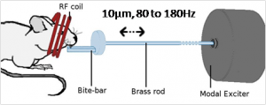
Brain MRE
In vivo Elastograms (storage and loss moduli ) of an anesthetized rat brain obtained by Low field MRE Mechanical excitation device on rat head for low field Magnetic Resonance Elastography
The quest for knowledge of the biomechanical behavior of brain tissue has been an essential issue for several decades. Determining the mechanical properties of brain tissue is an essential challenge in numerical modeling of the head.
Before to specify mechanical laws for finite element modeling, rheological properties of brain
tissue are needed. The results obtained in this way come from two kinds of protocols:
- Most of the results have been obtained through in vitro experimental protocols using classical rheometric techniques
- However recent in vivo studies for brain tissue have been published that use Magnetic Resonance Elastography (MRE). This non invasive technique is based on the coupling of an intra-cerebral shear wave propagating device and a MRI system.
Low field (0.1T) Magnetic Resonance Elastography device
Ex vivo and in vivo characterization of soft tissues including their dynamic behaviour, microscopic components, heterogeneity and anisotropy investigation is an essential topic to develop analytical models and at least more realistic models of the human brain.